Brain Plasticity During Skill Acquisition: Bridging the Gap Between Animal Models and Human Research
Center for Lifespan Psychology & Lise Meitner Group for Environmental Neuroscience
In this collaborative project, which involves three Max Planck Institutes, we investigate experience-dependent brain plasticity during motor skill acquisition. By carrying out coordinated experimental research with humans and mice and by using multimodal neuroimaging methods, we seek to gain a more mechanistic understanding of human brain plasticity.
In everyday life, humans show skilled performance on many tasks such as grasping an object, using a tool, or riding a bike. Typically, such tasks are learned by practicing them repeatedly over extended periods of time until they can be performed fluently and with little effort. This process of skill acquisition is put into operation by experience-dependent plasticity, which refers to the brain’s capacity to form lasting and behaviorally relevant structural and functional changes in neural connections that allow individuals to adapt to changing environmental demands.
In humans, plasticity in the course of motor skill acquisition has been observed at the macroscopic level in the form of grey-matter volume changes in primary motor cortex. In line with the exploration–selection–refinement (ESR) model of brain plasticity (Lindenberger & Lövdén, 2019; Lövdén et al., 2020), a non-monotonic macroscopic pattern was observed, consisting of tissue expansion followed by renormalization (Wenger et al., 2017). According to the ESR model, several sets of competing neuronal microcircuits potentially capable of implementing the computations needed to execute the to-be-learned skill are widely probed early in learning, and eventually structurally altered. This phase of exploration is followed by phases of experience-dependent selection and refinement of reinforced microcircuits, which lead to the concomitant gradual elimination of novel structures associated with unselected circuits, presumably resulting in macroscopically observable renormalization of tissue volume.
Unfortunately, it is not yet possible to study changes at the cellular level non-invasively in humans. Thus, to further validate the predictions of the ESR model, there is a dire need to map insights from microscopic measurements in animals onto macroscopic findings obtained in humans. The closer alignment of these two strands of research requires a systematic overlap in methodology across species. To accomplish this goal, researchers at the MPI for Human Development (MPIB) in Berlin, the MPI of Psychiatry (MPIP) in Munich, and the MPI for Biological Intelligence (MPIBI) in Martinsried near Munich have teamed up to coordinate their complementary expertise. Researchers at MPIB are well-versed in conducting human longitudinal studies investigating brain plasticity at the macroscopic level using magnetic resonance imaging (MRI). Researchers at MPIP obtain high-resolution anatomical brain scans from mice using a small-animal MRI system to identify brain structures involved in learning processes. Researchers at MPIBI are experts in investigating neuronal plasticity using in-vivo two-photon microscopy. The coordinated use of macroscopic and microscopic methods in humans and mice can provide new insights about the relationship between macroscopically observable volume changes and cellular processes in the context of experience-dependent brain plasticity.
Collaboration Partners / Network


One challenging precondition for the successful implementation of the project was to establish analogous behavioral protocols that are likely to elicit brain plasticity in both mice and humans. This requires explicit efforts to synchronize and adjust the learning tasks for humans and mice to enable comparable observations across species. We decided to set up protocols for motor skill acquisition, given that the behavioral assessment of a motor task and the localization of expected brain plasticity in motor regions are relatively straightforward in both species.
Both rodents and humans are capable of acquiring motor skills in the form of complex grasping movements. In mice, we used the well-established single-pellet reaching task to train skillful forelimb movements over several days. In this task, mice are trained to grasp a millet seed by reaching through a narrow slit and retrieve it to their mouth. We adapted this training regime to humans by designing a reaching task with chopsticks, in which participants need to grasp a single M&M (i.e., a chocolate-coated peanut) with chopsticks, transport it, and put it down in a bowl. Thus, both mice and humans learn to reach for and grasp a small food item that needs to be transported over a short distance.
In the main study, human participants trained the grasping task with chopsticks for 70 days in total. Each day, they needed to successfully grasp and transport 30 M&Ms. During the training period, participants were invited to the Institute on seven occasions to obtain structural and functional MR measurements. In the companion experiment in Munich, mice were trained to grasp 30 millet seeds for 25 days and underwent structural MRI on three occasions.
Another prerequisite for the project is to coordinate analysis pipelines of brain data suitable for rodent and human brains. Usually, in humans, the analysis of volume changes requires the segmentation of the brain into different tissue classes using established toolboxes. Segmentation of mice brains is generally more difficult, reflecting differences in image contrasts and less clearly defined brain structures (e.g., absence of cortical gyrification). To establish common ground across species, we searched for processing pipelines that can be applied to both human and mice brains such that comparisons across species are warranted. Currently, we are using deformation-based morphometry in addition to voxel-based morphometry; the former method has the advantage that it does not depend on brain segmentation.
The two motor tasks resulted in comparable learning curves across species (Figure 1). Initially, human participants took about 16 minutes to grasp 30 M&Ms with chopsticks. Within the first four weeks of training, there was a steep decrease in the time needed, followed by stable asymptotic performance in most participants. Specifically, participants’ performance reached a mean duration of about 5 minutes and 23 seconds to grasp 30 M&Ms on Day 30. Mice showed a similar decrease in time required to successfully grasp 30 millet seeds, starting with a mean duration of 14 minutes on day 1 and stabilizing their grasping time around day 8, with a mean duration of 4 minutes and 10 seconds.
 method.")
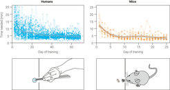
Figure 1. Performance improvements in the grasping tasks during the learning period. For each participant, individual data points of the learning curve are shown. A fitted line, smoothing the data series in each species, is depicted using the LOESS (locally estimated scatterplot smoothing) method.
Currently, project members are performing morphometric brain analyses with the acquired brain data from humans and mice to investigate brain changes over time during motor skill learning at the macroscopic level. Further analysis will entail two-photon microscopy processing of mouse brain data and estimation of microstructural tissue properties in human brains using MRI-based in-vivo histology.

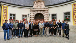
Figure 2. Participants in the Ringberg conference.
From left to right: Ju Lu, Yi Zuo, Constance Scharff, Amos Pagin, Andreas Lüthi, Jörn Diedrichsen, Manfred Gahr, Martin Lövdén, Iris Odstrcil, Tibor Stark, Daniel Vosberg, Warsha Barde, Geoffrey Delamare, Mallar Chakravarty, Maike Hille, Jason Lerch, Kristen Delevich, Adam Hantman, Elisabeth Wenger, Sara Zocher, Frederic Ullén, Maria Spolidoro, Robert Zatorre, Mark Hübener, Tomáš Paus, Claudia Buss, Takao Hensch, Nora Moog, Gerd Kempermann, Agnieszka Kulesza, Tobias Bonhoeffer, Simone Kühn, Ulman Lindenberger, Sandra Schmidt
Not in the photo: Pico Caroni, Claudia Clopath, Ania Majewska, Miriam Mosing, Tomás Ryan, Daniela Vallentin, Linda Wilbrecht
Ringberg Conference: Understanding Neural Plasticity: From Animal Models to Human Individuality
Inspired by this collaborative project, Tobias Bonhoeffer (MPIBI), Simone Kühn (MPIB), and Ulman Lindenberger (MPIB) organized a conference to discuss ways of bridging the gaps between animal models of brain plasticity and human research. The conference included 40 participants from 7 different countries and took place in September 2022 at Castle Ringberg, Bavaria. The conference fostered close interactions among researchers studying brain plasticity in different species and at different levels of analysis, and helped to generate new research ideas and experimental paradigms to advance comparative research on brain plasticity.
Research Project in Brief
Topic: Training-induced brain changes during motor skill learning in humans and mice
Researchers: Tobias Bonhoeffer (Director, Synapses – Circuits – Plasticity, MPI for Biological Intelligence), Michael Czisch (Head, Core Unit Neuroimaging, MPI of Psychiatry), Maike Hille (Predoctoral LIFE Fellow, Center for Lifespan Psychology & Lise Meitner Group for Environmental Neuroscience, MPI for Human Development), Lena Justus (Postdoc, Synapses – Circuits – Plasticity, MPI for Biological Intelligence), Simone Kühn (Head, Lise Meitner Group for Environmental Neuroscience, MPI for Human Development), Ulman Lindenberger (Director, Center for Lifespan Psychology, MPI for Human Development), Tibor Stark (Postdoc / Staff Scientist, Core Unit Neuroimaging, MPI of Psychiatry; now at Department Emotion Research, MPI of Psychiatry), Sarah Zocher (Postdoc, Synapses – Circuits – Plasticity, MPI for Biological Intelligence; LIFE Alumna; now at DZNE Dresden).
Period: 2019–ongoing
Funding: Max Planck Society
Selected publications: